
Introduction
Training frequency is an important variable to consider when designing a training program. This essentially refers to how often you train a muscle group and it’s often measured in a weekly basis – i.e. 1x/week, 2x/week, etc. The typical bodybuilding practice is to train each muscle group twice per week and, frankly, that’s a pretty decent practice that is probably going to work for most people. However, is it optimal for all muscle groups? Let’s check it out.
Training Frequency in Research
When it comes to analyzing training frequency in the literature, it’s pretty hard to come to a decent conclusion for a couple of reasons:
- Most research training programs are crap compared to actual bodybuilding or strength and conditioning programs.
- When researchers attempt to isolate frequency as a variable, they often equate weekly volume between different frequencies. With this in mind, a group training once per week might perform 4×10 on an exercise whereas the group training twice per week would just perform 2×10 on an exercise on two separate days. While this is great for investigating frequency, it defeats the whole purpose of increasing training frequency.
With these shortcomings, research has still mostly determined that training each muscle group twice per week is a little more effective than training each muscle group once per week (46). However, the confidence with which we can make these claims (from a scientific basis anyways) is lacking as this claim is based off of fewer than 10 studies with mostly untrained subjects.
Since research into training frequency isn’t helping us much, how else can we determine the optimal training frequency for each muscle group?

The Nitty Gritty
As bodybuilders or powerlifters, we often like to think that more is better. More weight, more reps, more protein, more sleep… the list never ends. So why wouldn’t more frequency be better? Do we have a good reason to not train every muscle group every day?
The goal of a training program is to provide progressive overload to each muscle group in order to make gains. However, each workout you perform is going to cause some level of muscle damage and soreness. This damage is caused by eccentric contractions (1,9) as well as biochemical disturbances that further damage the muscle fiber (5). Your soreness can then be exacerbated by the inflammation response to said muscle damage (9).
 So, what’s the problem with muscle damage? When our muscles are damaged, they cannot produce as much force as they can when they’re fresh (11,29). This means that you won’t be able to lift as much weight which leads to you not being able to progressively overload if you’re sore all of the time. In addition, your overall muscle activation is likely lower when you’re sore (39) which will reduce the amount of muscle fibers that get a growth stimulus from that workout. Therefore, there’s really no point in training while you’re sore as you won’t make much (if any) progress, and you likely have a greater chance of injury (16).
With the damage and soreness issue in mind, training frequency should probably be based off of how easily a muscle group is damaged and how quickly it can recover from that damage. Interestingly enough, just about every muscle will recover at a different rate (11), meaning that each muscle might require a different training frequency. Since frequency should be predicated on damage and recovery, what factors influence damage and recovery?

Muscle Size
The size of a muscle is one of the first places we look to determine how much recovery time it might need between sessions and, therefore, how often you can train it. Now, some might assume that larger muscles require greater recovery periods simply due to their size but it’s a little more complex than that. The first item that throws a wrench in that wheel of thought is the fact that the eye test isn’t always accurate for muscles – i.e. many would assume that the lats are a large muscle group, but the triceps, pecs, and deltoids are actually much larger by muscle volume (28). So, now we have to dig deeper.
Regardless of what your girlfriend tells you, size does matter. But size influences recovery a little differently than many would think. Typically (but not always), muscle size will influence how well you can activate that muscle. This is reflected in the findings showing that small muscles, like the biceps, are very easily activated (15) compared to larger muscles, like the quadriceps, that don’t activate nearly as well (4,6). This size test isn’t 100% accurate since the triceps are activated just as well (if not more so) than the biceps (12), but we have limited information on many muscle groups when it comes to activation. When we don’t have published activation levels, we can use the size of the muscle to theorize how well we can activate it since we have plenty of info on muscle volume – more about that at the end of this article.
Anywho, why does activation matter? Activation tells us how many fibers are active in a given contraction – this is typically expressed as a % of a maximal voluntary or involuntary contraction. Like we discuss above, most healthy people can voluntarily activate their biceps in excess of 95% of a maximal involuntary contraction, which requires electrical stimulation (4,15). On the other hand, subjects often show voluntary activation levels in the 80-85% range for the quadriceps when compared to involuntary contractions (4).
So, if a muscle is harder to activate, how would that influence recovery? If you have less voluntary activation, you have fewer active fibers. And, if you have fewer active fibers, you won’t get as much muscle damage. And, to put it all together, less muscle damage leads to less recovery time required. Therefore, we can typically train larger muscle groups with more frequency than smaller muscle groups, but there’s a few more caveats to follow before we can really finalize that claim.
Muscle Fiber Type
The next topic to discuss that might have the most influence on training frequency for a given muscle is the predominant fiber type of that muscle. Why does this matter?
Remember, we’re mostly basing training frequency off of muscle damage and subsequent recovery time. With this in mind, it’s important to understand that fast twitch muscle fibers will typically experience more muscle damage than slow twitch fibers (22). This is mostly due to these fibers being less oxidative in nature (biochemical stress) and producing more force in general (mechanical stress). We see further proof of this scenario when comparing protein synthesis rates between fast and slow twitch muscle fibers following training. Fast twitch fibers generally have a greater protein synthesis rate (23,32) due to increased levels of damage and greater repair needs.
Therefore, muscle groups that have a higher percentage of fast twitch muscle fibers will probably need more recovery time between training sessions and, thus, should be trained less frequently than muscles with more slow twitch fibers.
We just about have the theoretical background set for training frequency, but there’s one more component worth discussing.
Joint Range of Motion
Joint range of motion is the last place to look as this can influence the amount of length change and loaded stretch that a muscle will undergo. Now, some of the more astute readers may have noticed that our recommendations are very similar to Dr. Chris Beardsley’s (website here) when it comes to training frequency, but this last point is where we differ slightly.
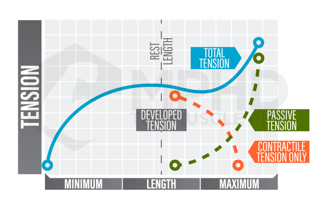 Dr. Beardsley discusses sarcomeres and their relation to the length-tension curve as his last component of frequency. If that sound like gibberish to you, don’t worry. Sarcomeres are essentially the contractile units of muscle – these are the individual units that shorten or lengthen to cause a muscle contraction. Now, the sarcomeres within specific motor units of each muscle group are generally assigned to one portion of the length-tension curve. Some sarcomeres may only be active on certain portions of the curve whereas others are active on different portions of the curve. The descending limb (right of center) of the curve is typically associated with a greater stretch and greater overall tension on the muscle fiber.
Some sarcomeres can actively reach this range without issue; however, not all sarcomeres share that ability. The sarcomeres that are only active on the ascending and/or plateau portions of the curve (left of center) are more likely to be damaged from full range of motion movements (8) which would obviously increase recovery needs. With this in mind, knowing information about sarcomere length-tension relationships in each muscle group can help determine frequency.
Unfortunately, further investigation of this presents some issues. We don’t have information on all muscle groups (we’re currently compiling as much as possible for a new article). For other muscle groups, it gets a little muddy when you compare muscles that perform the same action – like the soleus and gastrocnemius, for instance. The soleus does have sarcomeres that operate on all portions of the length-tension curve (10) whereas the gastroc is limited to just the ascending portion (36). Therefore, since we train both muscles with calf raises, this knowledge doesn’t really do much for us in the long run when it comes to frequency.
So, our team has come up with a slightly more applicable method to help finalize determination of the optimal training frequency for each muscle group – joint range of motion.
No matter the sarcomere length-tension relationship of a given muscle, the factor that is going to induce stretch (and potential for damage) is the amount of range of motion a joint can go through – with greater ROMs inducing more damage (42). We see the importance of joint ROM when comparing hypertrophy adaptations to full squats and partial squats – full squats obviously result in greater overall muscle gain in the quads (7,33), adductors, and glutes (33). This is due to a general increase in mechanical tension from the greater length change that the full range of motion causes. While it’s a bit of a generalization, performing exercises that require a greater range of motion will likely increase muscle damage (34,42).
With this in mind, the muscles that cross joints with a larger ROM (such as the shoulder) usually have a wide selection of large ROM exercise variations. Interestingly enough, these muscles are also usually more fast twitch in nature and likely already require more recovery time, but the increased ROM of the shoulder joint in certain exercises can easily play a role in stretch-induced damage. Therefore, when we discuss frequency, we also need to consider what exercises you’re doing and what kind of ROM you’re putting the muscle through.
Since we’ve now laid out how you can start planning frequency, let’s now cover these topics by muscle group so we can make some applicable takeaways. We’ll discuss the size (and voluntary activation when applicable), muscle fiber type, and joint ROM considerations to determine the optimal frequency for each muscle.
Training Frequency by Muscle Group
Calves
Obviously, we have to start with the most important muscle group. We cover the calves in more detail in our Calf Training 101 article (here) if you’re really intrigued (we know you are…).
Muscle Size: The calves certainly aren’t the biggest muscle in the body, but they are definitely bigger than the biceps (13) unless you have absolute twigs for legs. However, their voluntary activation levels are actually pretty high (98%+) (13) so they’re one of the sneaky muscles that can throw off the size trick a little bit.
Muscle Fiber Type: The calves are very well-known to be mostly slow twitch, with the gastroc presenting up to 76% slow twitch (14) and the soleus having a range of 70-96% slow twitch (14).
Joint ROM: The calf muscles are primarily stretched when the ankle is in dorsiflexion – i.e. when the toes are pointing up. The degree of this stretch can influence damage and recovery times, but the reality is that this range of motion is much shorter than many other joints (38). Therefore, to optimize tension on the calves, using strategies that emphasize what little stretch you can get in the ankle joint might help boost growth – however, it will likely increase recovery time, too.
Conclusion: Due to their extreme slow twitch dominance and generally small joint ROM, the calves can probably be trained quite frequently. In support of this, studies show that the calves are just about tied with the quads for the fastest recovery rates (11). Therefore, aim to add some sort of calf training to workouts 4-5 days/week – AKA to all of your workouts, for most of you. This can be easily accomplished by supersetting other exercises with calf raise variations throughout the week – for example, you’ll be resting 2-3 minutes between sets on bench, might as well add some calf raises during this time!
Quads
 We’ve already touched on the quads quite a bit as they’re a great example of many of the theories behind frequency. Regardless, we’ll still sum up the findings here.
Muscle Size: The quads are easily one of the largest muscle groups in the body (17,41) and we already know that their voluntary activation levels are quite low (4).
Muscle Fiber Type: This can get a little tricky as we do have 4 separate muscles within our quadriceps and they do present with some different fiber type compositions. The largest quadriceps muscle is the vastus lateralis (17) and it’s typically right about a 50-50 split between fast twitch and slow twitch (25). However, other quad muscles, like the rectus femoris, are slightly more fast twitch oriented (48) and might get damaged a little more easily.
Joint ROM: The quadriceps are primarily knee extensors, so we need to inspect knee flexion ROM to assess how this might stretch the quads. The knee can achieve rather large degrees of flexion (38) meaning that deeper squats or lunges will likely cause a little more damage to the quads. Therefore, using full ROM movements might increase recovery time in the quads, but we can still expect the quads to recover rather quickly.
Conclusion: Due to their massive size and poor level of voluntary activation, the quads can also be trained rather frequently. Like we stated above, the quads and calves are the quickest muscle groups to recover following damaging training (11), however, it’s important to keep in mind that the quads are highly involved in movements like squats and lunges. Squats and lunges are also going to cause muscle damage to muscle groups like the adductors that don’t recover as quickly as the quads. Therefore, even though you should train your quads pretty often, you shouldn’t do so by adding more squats or lunges throughout the week. I think 2-3 big leg days per week is fine, but then add some quad isolation work to another 1-2 workouts per week to hit the quads 4-5 days per week. This can be accomplished by supersetting other exercises with things like leg extensions or sissy squats.
Hamstrings
In case you haven’t noticed, we’re mostly taking a ground-up approach here. Therefore, the next logical step for discussion is the hamstrings group.
Muscle Size: We don’t have a ton of data on hamstrings muscle volume as the hamstrings group also technically consists of 3 muscles and 4 total muscle heads. Regardless, the hamstrings are certainly smaller than the quads (20,40) and can achieve a much higher voluntary activation level (~98%) (2).
Muscle Fiber Type: Many seem to think that the hamstrings are mostly fast twitch muscles, and in elite athletes, this is probably true. However, for the other 99.99% of humans, the hamstrings are a pretty even 50-50 split (19).
Joint ROM: The hamstrings are a biarticular muscle group (except for the short head of the biceps femoris) and can be stretched at both the knee and the hip, during extension and flexion, respectively. The knee obviously is only going to have a few degrees of hyperextension, so this alone won’t stretch the hamstrings much. However, the hip has a pretty large ROM when considering hip flexion (38) which can absolutely stretch the hammies – especially near full knee extension.
Conclusion: Due to extremely high levels of voluntary activation, the hamstrings are probably going to be decently damaged from exercise – especially movements that involve large degrees of hip flexion with mostly extended knees (stiff leg deadlifts, etc.). This is reflected in the findings that the hamstrings are about middle-of-the-pack when it comes to recovery rates for muscle groups (11). Therefore, I wouldn’t train the hamstrings as often as the quads or calves, but they can still be trained a little more often than many upper body muscles. I’d shoot for 3-days a week on hamstrings; you might be able to squeeze out a fourth day if you add in primarily knee flexion exercises that won’t cause as much stretch or damage.
Glutes
Continuing our ground-up approach, let’s satisfy Sir Mix-a-Lot and talk about booty frequency.
Muscle Size: The gluteal group is well-known to be the largest muscle group in the human body (49) with the gluteus maximus being the largest single muscle in the body (44). However, we have very little data on their voluntary activation levels. We do know that training can improve this level of voluntary activation (21), but that’s a very common neuromuscular adaptation to training that would occur in any muscle group. Regardless, it might suggest that the glutes are a little lower than other muscle groups since they obviously have significant room to improve.
Muscle Fiber Type: We don’t have a ton of data on glute fiber types in healthy individuals. The one study we do have shows the glutes to be slightly more slow twitch than fast twitch (30) but this could certainly vary between individuals and even regions of the glute muscles themselves.
Joint ROM: The glutes primarily act on the hip through a variety of actions. The main way the glutes would be stretched is through hip flexion, and we already know from our hamstrings discussion that the hip flexion ROM is pretty large (38). Therefore, movements that involve more hip flexion might increase recovery time for the glutes.
Conclusion: We don’t have much concrete data on the glutes, but from their size alone I think we can theorize that you can get away with training the glutes rather often. Again, this will be a case similar to the quads in which you’ll have to add isolation exercises to other workouts in order to increase this frequency. I think 2-3 big leg days per week is fine and you can likely add glute isolation work to 1-2 other workouts throughout your week. Squats and lunges will likely damage the glutes more, but you can add in machine work or hip thrusts for your additional glute days throughout the week.
Core
We’ll label this section “core” as most core muscles will have a similar fiber type percentage and ROM. However, size can certainly vary more, but this is just an article and not a dissertation so we won’t go into every single core muscle individually. We’ll mainly discuss the rectus abdominis (RA) since it’s the 6-pack muscle that every bodybuilder seeks to develop.
Muscle Size: The RA is a rather large muscle compared to most upper body muscles (45). This size does influence muscle activation as the RA has been shown to have pretty low voluntary activation (86%) (18) which makes it nearly as hard to activate as the quads.
Muscle Fiber Type: RA is mostly slow twitch (26) due to its involvement with spine stabilization throughout the day. Certain neuromuscular compartments of various core muscles might be a little more fast twitch as they are used for things like rotation, etc. but we have little to no data on that theory.
Joint ROM: RA is going to be active in multiple planes of movement so it’s a bit short-sighted to isolate joint ROM to spine flexion. Regardless of this freedom, RA doesn’t go through a large range of motion in any plane and isn’t exactly designed to lengthen to a great degree anyways (37).
Conclusion: For just about every reason listed, the abs (especially RA) can probably be trained pretty often. The abs recover nearly as quickly as the quads and calves (11) so you’re likely fine training abs about 4-days a week. Just make sure to plan ab training around heavy compound movements so that your abs aren’t fatigued when you’re trying to hit heavy squats or deadlifts.
Lats
 Alright, alright, we’re officially on to the classic upper body muscles. First up, the lats.
Muscle Size: Like we mentioned in the beginning of this piece (feels like forever ago, right?), the lats are kind of an ambiguous muscle. From the outside, they look massive, but realistically, they’re a pretty thin muscle and are actually quite a bit smaller than muscles like the deltoids, triceps, and pecs (28). We don’t have any data (that we know of) considering the voluntary activation % of the lats, but we can assume it to be decent since even larger upper body muscles, like the triceps, can still achieve a high level of activation (12).
Muscle Fiber Type: The lats are another muscle group we don’t have a ton of info on (you’re probably seeing a trend here – beyond quads and calves, we don’t have much data). However, some studies suggest the lats are close to a 50-50 split whereas others suggest they might be slightly fast twitch dominant (43).
Joint ROM: The lats attach to the upper arm and cause actions at the shoulder joint. The shoulder can undergo massive ranges of motion which means that the lats can be placed under stretch in a variety of exercises. Emphasizing the stretch in pulling exercises (especially pulldowns) might require additional recovery time for the lats.
Conclusion: The lats can probably be activated pretty well and might be slightly fast twitch – plus, they can undergo huge ranges of motion since they act on the shoulder joint. These findings are probably why the lats are a little slower to recovery from training (11) but they do still beat out some other upper body muscle groups. I’d stick to 3x/week max for training lats – if you do some shorter range of motion work, you might be able to squeeze out an additional isolation day each week.
Traps
Since we’re already on the back half of the body, let’s round out the major back muscles with a discussion on the traps.
Muscle Size: The traps are another muscle group that appears pretty large, but they’re actually a little smaller than the lats (~230cm3 vs ~260cm3) (50). With this smaller size in mind, the traps have a pretty high voluntary activation level (~95%) (3).
Muscle Fiber Type: Fiber type will vary somewhat between the different segments of the traps, but for the most part, the traps have a pretty high percentage of slow twitch fibers (66-80% in the middle and lower traps) (35). The upper traps are nearly a 50-50 split, but this segment of the traps represents the smallest and least active portion of the traps compared to the middle and lower traps. Contrary to popular belief, the middle traps are the most responsible for shrugging motions than the upper traps, but that’s a conversation for another time.
Joint ROM: The traps primarily act on the scapula (shoulder blade) and it can be somewhat difficult to quantify range of motion at the scapula. This can be done by assessing degrees of rotation during pulldown exercises but rotation measurements for protraction and retraction are a little more complex. Regardless of these difficulties, we do know that maintaining a more upright posture during rowing and pulldown movements can help increase the range of motion the scapula goes through during the exercise (31). This could slightly influence the stretch that the traps undergo during these movements, but it still likely won’t be that large. In addition, the scapulae will undergo a decent range of motion during shrugging exercises (27) so there’s a slight stretch if you hold the bottom of a shrug, but it’s still not as much as many other muscle groups.
Conclusion: Mostly due to their highly slow twitch nature, the traps can probably be trained very often. I think this group can get lumped in with the quads and calves for being trained 4-5x/week. However, like the quads, it’s important to keep in mind that movements that train the traps can also damage other muscle groups – like rows and pulldowns. Therefore, your additional trap workouts should mostly consist of isolation work like shrugs or even upright rows.
Pecs
Man, we almost saved the best for last. How mad would you have been if we skipped the pecs entirely?
Muscle Size: The pecs are slightly smaller in volume than the triceps (28) but I don’t think we have any data on their voluntary activation. Going off of size alone, we can probably theorize that the pecs slot somewhere between the triceps and biceps and are likely in the 95-98% range.
Muscle Fiber Type: Again, not much data here (shocker) but we do have one study showing that the pecs consist of 65% fast twitch fibers (47). This makes the pecs one of the fastest, if not the fastest, muscle groups in the human body.
Joint ROM: Like we’ve discussed a few times already, the pecs mainly act on the shoulder joint which can undergo a massive range of motion. This means that the pecs can be stretched quite a bit – especially in movements like wide grip bench press, dumbbell flyes, cable flyes, etc.
Conclusion: As you can see, the pecs are easily the muscle group that should be trained with the least frequency. This theory is supported by the finding that the pecs are the slowest muscle group to recover following training (11). I think 2x/week for chest training is plenty as the pecs will need at least a few days to recover between sessions.
Arms
It’s almost like we saved the most exciting muscle groups for last for a reason… time to discuss arms!
Muscle Size: We’ve used both the biceps and triceps as examples throughout this piece multiple times so we can simply recap here. The triceps are certainly larger than the biceps (28) but both muscles are able to achieve at least 95% of voluntary activation.
Muscle Fiber Type: Both the biceps and triceps are mostly fast twitch with the biceps hanging around 62% and the triceps consisting of about 57% fast twitch fibers (47). They’re not quite as fast twitch dominant as the pecs, but they’re certainly more fast twitch than many other muscle groups.
Joint ROM: Both muscles cross the elbow joint, but the elbow can obviously flex through a much larger motion than it can hyperextend (38). Therefore, the triceps will probably achieve more stretch during certain exercises than the biceps, but exercises like preacher curls can place a little more stretch on the biceps.
Conclusion: Due to all of the above, the arms probably should be trained less frequently than most lower body muscles, but possible a little more than the pecs. I think you can get away with training biceps more often than triceps since the elbow doesn’t undergo as great of a ROM during biceps exercises compared to triceps. Therefore, I think triceps should be hit 2-3x/week and you might be able to get an extra workout out of the biceps throughout the week for a frequency of 3-4x/week. Personally, I’ve had great results adding 3-5 sets of biceps to my usual 4-5 workouts per week rather than training them for 8-10 sets twice per week. Just an anecdote, but worth trying!
Conclusion
If you made it this far, congratulations! We know this piece was a long one but, frankly, we’re just not satisfied with the current state of the literature regarding training frequency. Training frequency can be much more intricate than just, “hit each muscle group 2x/week and you’re fine.” While that could certainly work for most muscles, it’s just too simple (and boring) for us at Team Muscle PhD to reiterate.
With the recommendations in this piece, you might be wondering how the hell you’re supposed to design a training split now. Add calves, traps, quads, and glutes 4-5x/week? This is where you need to get creative. Which one of these muscles are you really wanting to bring up right now? Let’s just assume it’s your calves since it probably is. Therefore, your calves get first dibs on this training cycle. Add calves into your workouts 4-5 days a week for a month or two, then dial them back to a maintenance 2x/week plan. When you dial calves back, move one of the other muscle groups into that 4-5x/week slot. Rotate these muscles groups every month or two so that you’re still capitalizing on their recovery capacity, but you’re not turning all of your workouts into 3-hour sessions.
As always, we’d love to see more research that analyzes training frequency by muscle group more specifically. We don’t have much yet, but I think the Chen et al. (2019) (reference #11) is a great start towards going down that road. If and when we get more info, we’ll be sure to update this piece.
Easter Egg
Since we have enough data, we’ll include a chart so you can get a better idea of the size of each muscle group relative to one another. This can help you somewhat theorize activation levels for muscles we don’t have data on. Keep in mind that results are coming from multiple studies and measuring techniques will be slightly different between research groups. I think this list is still a good general proxy of muscle size, however. With this being said, to ensure the data was as consistent as possible, we had to take a few steps:
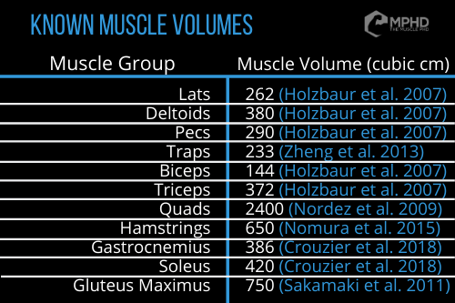 Most of these studies had both male and female subjects so we took the average volume between males and females in each study for the reported number on the chart.
- One study (Sakamaki et al. 2011) utilized a training program and published gluteus maximus volume before and after the training intervention. Since we’re attempting to keep data consistent here, we used the pre-training data for the glute volume on our chart.
- There are a few other studies out there that compare muscle volumes in various types of athletes, but we wanted to stray away from those and lean towards untrained subjects. We feel that an untrained population might better represent muscle volumes relative to one another. Commonly researched athletes, like soccer players, might have much more developed lower bodies than upper bodies which would throw things off a bit.
- The quads are also published as a collective rather than each individual quadriceps muscle so that one might not be as accurate (but saves space – hamstrings are also shown as a collective). This is why the quads appear much larger here – individual quad muscle volumes in untrained subjects are as follows (Ema et al. 2017): Vastus Medialis – 403cm3; Vastus Lateralis – 520cm3; Vastus Intermedius – 455cm3; Rectus Femoris – 228cm3. Trained rowers presented with muscle volumes of (Ema et al. 2017): Vastus Medialis – 532cm3; Vastus Lateralis – 798cm3; Vastus Intermedius – 608cm3; Rectus Femoris – 296cm3.
- To our knowledge, no published data exists on muscle volumes in elite bodybuilders. We’ll keep an eye out for that. Or we’ll do it ourselves, who knows….
References
- Allen, D. G. (2001). Eccentric muscle damage: mechanisms of early reduction of force. Acta Physiologica Scandinavica, 171(3), 311-319.
- Baumert, P., Temple, S., Stanley, M. J., Cocks, M., Strauss, J. A., Shepherd, S. O., … & Erskine, R. M. (2019). Neuromuscular fatigue and recovery after strenuous exercise depends on skeletal muscle size and stem cell characteristics. BioRxiv, 740266.
- Bech, K. T., Larsen, C. M., Sjøgaard, G., Holtermann, A., Taylor, J. L., & Søgaard, K. (2017). Voluntary activation of the trapezius muscle in cases with neck/shoulder pain compared to healthy controls. Journal of Electromyography and Kinesiology, 36, 56-64.
- Behm, D. G., Whittle, J., Button, D., & Power, K. (2002). Intermuscle differences in activation. Muscle & Nerve, 25(2), 236-243.
- Belcastro, A. N. (1993). Skeletal muscle calcium-activated neutral protease (calpain) with exercise. Journal of Applied Physiology, 74(3), 1381-1386.
- Beltman, J. G. M., Sargeant, A. J., Van Mechelen, W., & De Haan, A. (2004). Voluntary activation level and muscle fiber recruitment of human quadriceps during lengthening contractions. Journal of Applied Physiology, 97(2), 619-626.
- Bloomquist, K., Langberg, H., Karlsen, S., Madsgaard, S., Boesen, M., & Raastad, T. (2013). Effect of range of motion in heavy load squatting on muscle and tendon adaptations. European Journal of Applied Physiology, 113(8), 2133-2142.
- Brockett, C. L., Morgan, D. L., Gregory, J. E., & Proske, U. (2002). Damage to different motor units from active lengthening of the medial gastrocnemius muscle of the cat. Journal of Applied Physiology, 92(3), 1104-1110.
- Clarkson, P. M., & Hubal, M. J. (2002). Exercise-induced muscle damage in humans. American Journal of Physical Medicine & Rehabilitation, 81(11), S52-S69.
- Chen, X., & Delp, S. L. (2016). Human soleus sarcomere lengths measured using in vivo microendoscopy at two ankle flexion angles. Journal of Biomechanics, 49(16), 4164-4167.
- Chen, T. C., Yang, T. J., Huang, M. J., Wang, H. S., Tseng, K. W., Chen, H. L., & Nosaka, K. (2019). Damage and the repeated bout effect of arm, leg, and trunk muscles induced by eccentric resistance exercises. Scandinavian Journal of Medicine & Science in Sports, 29(5), 725-735.
- Cheng, A. J., & Rice, C. L. (2010). Voluntary activation in the triceps brachii at short and long muscle lengths. Muscle & Nerve: Official Journal of the American Association of Electrodiagnostic Medicine, 41(1), 63-70.
- Crouzier, M., Lacourpaille, L., Nordez, A., Tucker, K., & Hug, F. (2018). Neuromechanical coupling within the human triceps surae and its consequence on individual force-sharing strategies. Journal of Experimental Biology, 221(21).
- Dahmane, R., Djordjevič, S., Šimunič, B., & Valenčič, V. (2005). Spatial fiber type distribution in normal human muscle: histochemical and tensiomyographical evaluation. Journal of Biomechanics, 38(12), 2451-2459.
- De Serres, S. J., & Enoka, R. M. (1998). Older adults can maximally activate the biceps brachii muscle by voluntary command. Journal of Applied Physiology, 84(1), 284-291.
- Dupuy, O., Douzi, W., Theurot, D., Bosquet, L., & Dugué, B. (2018). An evidence-based approach for choosing post-exercise recovery techniques to reduce markers of muscle damage, soreness, fatigue, and inflammation: a systematic review with meta-analysis. Frontiers in Physiology, 9, 403.
- Ema, R., Wakahara, T., Hirayama, K., & Kawakami, Y. (2017). Effect of knee alignment on the quadriceps femoris muscularity: Cross-sectional comparison of trained versus untrained individuals in both sexes. PloS One, 12(8).
- Ertman, H., Szepietowski, O., Chiou, S. Y., & Strutton, P. H. (2016). Voluntary activation of abdominal muscles assessed using transcranial magnetic stimulation. Orthopaedic Proceedings, 98(6). The British Editorial Society of Bone & Joint Surgery.
- Evangelidis, P. E., Massey, G. J., Ferguson, R. A., Wheeler, P. C., Pain, M. T., & Folland, J. P. (2017). The functional significance of hamstrings composition: is it really a “fast” muscle group? Scandinavian Journal of Medicine & Science in Sports, 27(11), 1181-1189.
- Evangelidis, P. E., Massey, G. J., Pain, M. T., & Folland, J. P. (2016). Strength and size relationships of the quadriceps and hamstrings with special reference to reciprocal muscle balance. European Journal of Applied Physiology, 116(3), 593-600.
- Fisher, B. E., Southam, A. C., Kuo, Y. L., Lee, Y. Y., & Powers, C. M. (2016). Evidence of altered corticomotor excitability following targeted activation of gluteus maximus training in healthy individuals. Neuroreport, 27(6), 415-421.
- Friden, J. (1992). Structural and mechanical basis of exercise-induced muscle injury. Medicine and Science in Sports and Exercise, 16, 456-459.
- Goodman, C. A., Kotecki, J. A., Jacobs, B. L., & Hornberger, T. A. (2012). Muscle fiber type-dependent differences in the regulation of protein synthesis. PloS One, 7(5).
- Gondin, J., Guette, M., Maffiuletti, N. A., & Martin, A. (2004). Neural activation of the triceps surae is impaired following 2 weeks of immobilization. European Journal of Applied Physiology, 93(3), 359-365.
- Gouzi, F., Maury, J., Molinari, N., Pomiès, P., Mercier, J., Préfaut, C., & Hayot, M. (2013). Reference values for vastus lateralis fiber size and type in healthy subjects over 40 years old: a systematic review and meta-analysis. Journal of Applied Physiology, 115(3), 346-354.
- Häggmark, T., & Thorstensson, A. (1979). Fibre types in human abdominal muscles. Acta Physiologica Scandinavica, 107(4), 319-325.
- Hallaceli, H., & Günal, I. (2002). Normal range of scapular elevation and depression in healthy subjects. Archives of Orthopaedic and Trauma Surgery, 122(2), 99-101.
- Holzbaur, K. R., Murray, W. M., Gold, G. E., & Delp, S. L. (2007). Upper limb muscle volumes in adult subjects. Journal of Biomechanics, 40(4), 742-749.
- Johar, P., Grover, V., Topp, R., & Behm, D. G. (2012). A comparison of topical menthol to ice on pain, evoked tetanic and voluntary force during delayed onset muscle soreness. International Journal of Sports Physical Therapy, 7(3), 314.
- Johnson, M., Polgar, J., Weightman, D., & Appleton, D. (1973). Data on the distribution of fibre types in thirty-six human muscles: an autopsy study. Journal of the Neurological Sciences, 18(1), 111-129.
- Kebaetse, M., McClure, P., & Pratt, N. A. (1999). Thoracic position effect on shoulder range of motion, strength, and three-dimensional scapular kinematics. Archives of Physical Medicine and Rehabilitation, 80(8), 945-950.
- Koopman, R., Zorenc, A. H., Gransier, R. J., Cameron-Smith, D., & van Loon, L. J. (2006). Increase in S6K1 phosphorylation in human skeletal muscle following resistance exercise occurs mainly in type II muscle fibers. American Journal of Physiology-Endocrinology and Metabolism, 290(6), E1245-E1252.
- Kubo, K., Ikebukuro, T., & Yata, H. (2019). Effects of squat training with different depths on lower limb muscle volumes. European Journal of Applied Physiology, 119(9), 1933-1942.
- Lieber, R. L., & Friden, J. (1993). Muscle damage is not a function of muscle force but active muscle strain. Journal of Applied Physiology, 74(2), 520-526.
- Lindman, R., Eriksson, A., & Thornell, L. E. (1990). Fiber type composition of the human male trapezius muscle: Enzyme‐histochemical characteristics. American Journal of Anatomy, 189(3), 236-244.
- Maganaris, C. N. (2003). Force‐length characteristics of the in vivo human gastrocnemius muscle. Clinical Anatomy: The Official Journal of the American Association of Clinical Anatomists and the British Association of Clinical Anatomists, 16(3), 215-223.
- McGill, S. (2010). Core training: Evidence translating to better performance and injury prevention. Strength & Conditioning Journal, 32(3), 33-46.
- McKay, M. J., Baldwin, J. N., Ferreira, P., Simic, M., Vanicek, N., Burns, J., & 1000 Norms Project Consortium. (2017). Normative reference values for strength and flexibility of 1,000 children and adults. Neurology, 88(1), 36-43.
- Nie, H., Arendt-Nielsen, L., Kawczynski, A., & Madeleine, P. (2007). Gender effects on trapezius surface EMG during delayed onset muscle soreness due to eccentric shoulder exercise. Journal of Electromyography and Kinesiology, 17(4), 401-409.
- Nomura, Y., Kuramochi, R., & Fukubayashi, T. (2015). Evaluation of hamstring muscle strength and morphology after anterior cruciate ligament reconstruction. Scandinavian Journal of Medicine & Science in Sports, 25(3), 301-307.
- Nordez, A., Jolivet, E., Südhoff, I., Bonneau, D., de Guise, J. A., & Skalli, W. (2009). Comparison of methods to assess quadriceps muscle volume using magnetic resonance imaging. Journal of Magnetic Resonance Imaging: An Official Journal of the International Society for Magnetic Resonance in Medicine, 30(5), 1116-1123.
- Nosaka, K., & Sakamoto, K. E. I. (2001). Effect of elbow joint angle on the magnitude of muscle damage to the elbow flexors. Medicine & Science in Sports & Exercise, 33(1), 22-29.
- Paoli, A., Pacelli, Q. F., Cancellara, P., Toniolo, L., Moro, T., Canato, M., … & Reggiani, C. (2016). Protein supplementation does not further increase latissimus dorsi muscle fiber hypertrophy after eight weeks of resistance training in novice subjects, but partially counteracts the fast-to-slow muscle fiber transition. Nutrients, 8(6), 331.
- Sakamaki, M., Bemben, M. G., & Abe, T. (2011). Legs and trunk muscle hypertrophy following walk training with restricted leg muscle blood flow. Journal of Sports Science & Medicine, 10(2), 338.
- Sanchis-Moysi, J., Idoate, F., Dorado, C., Alayón, S., & Calbet, J. A. (2010). Large asymmetric hypertrophy of rectus abdominis muscle in professional tennis players. PLoS One, 5(12).
- Schoenfeld, B. J., Ogborn, D., & Krieger, J. W. (2016). Effects of resistance training frequency on measures of muscle hypertrophy: a systematic review and meta-analysis. Sports Medicine, 46(11), 1689-1697.
- Srinivasan, R. C., Lungren, M. P., Langenderfer, J. E., & Hughes, R. E. (2007). Fiber type composition and maximum shortening velocity of muscles crossing the human shoulder. Clinical Anatomy: The Official Journal of the American Association of Clinical Anatomists and the British Association of Clinical Anatomists, 20(2), 144-149.
- Stålberg, E., Borges, O., Ericsson, M., Essén‐Gustavsson, B., Fawcett, P. R. W., Nordesjö, L. O., … & Uhlin, R. (1989). The quadriceps femoris muscle in 20–70‐year‐old subjects: relationship between knee extension torque, electrophysiological parameters, and muscle fiber characteristics. Muscle & Nerve: Official Journal of the American Association of Electrodiagnostic Medicine, 12(5), 382-389.
- Ward, S. R., Eng, C. M., Smallwood, L. H., & Lieber, R. L. (2009). Are current measurements of lower extremity muscle architecture accurate? Clinical Orthopaedics and Related Research, 467(4), 1074-1082.
- Zheng, L., Siegmund, G., Ozyigit, G., & Vasavada, A. (2013). Sex-specific prediction of neck muscle volumes. Journal of Biomechanics, 46(5), 899-904.
From being a mediocre athlete, to professional powerlifter and strength coach, and now to researcher and writer, Charlie combines education and experience in the effort to help Bridge the Gap Between Science and Application. Charlie performs double duty by being the Content Manager for The Muscle PhD as well as the Director of Human Performance at the Applied Science and Performance Institute in Tampa, FL. To appease the nerds, Charlie is a PhD candidate in Human Performance with a master’s degree in Kinesiology and a bachelor’s degree in Exercise Science. For more alphabet soup, Charlie is also a Certified Strength and Conditioning Specialist (CSCS), an ACSM-certified Exercise Physiologist (ACSM-EP), and a USA Weightlifting-certified performance coach (USAW).